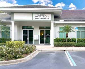
October 15, 2008
How To Install a RAID Configuration on Windows XP
Here is a step-by-step guide on how to install a RAID configuration on Windows XP. I would encourage anyone contemplating this to read as many tech forums on this subject as is reasonable.
Read Steps 1-6 BEFORE PROCEEDING:
1. Build your computer with all the components installed including the drives for RAID configuration. If you have a card reader, unplug this from the MOBO port for now. Start the computer and make sure that all fans are running and you can boot into BIOS. If you already have a prebuilt computer, do the same. Sorry, you WILL have to reinstall Windows XP. BACK UP YOUR DATA FIRST. ONCE THE OS IS INSTALLED, YOU CANNOT GO BACK AND INSTALL RAID DRIVES. THE RAID INSTALL MUST OCCUR PRIOR TO AND DURING OS INSTALLATION. You will also need a REGULAR (NO USB) FLOPPY DRIVE hooked into your MOBO. If necessary just scavenge one from an older computer and hook it up temporarily. Vista users can use USB. It would be best to flash your BIOS to the most current stable version.
2. Boot into your BIOS. The go to Main - SATA Configuration and change the setting from IDE to RAID. Hit Enter. Go to Boot Sequence and make sure that the CD/DVD drive is the first boot device. Hit Enter then F10 to Save and Exit. Reboot.
3. At the POST, watch for the RAID Screen, it will only be there for 3 seconds. Hit <CTRL – I> to enter the RAID configuration screen. From there select “Create RAID volume”, name it with no spaces. Select RAID type (0, 1, 5 or 10), Select drives to be included (don’t include your main drive (C:) for RAID 5), Select striping rate (64 or 128) Hit Enter. Create RAID volume. ENTER and exit.
4. On another computer, that has a floppy drive. Go to Intel’s website and search for Intel® Matrix Storage Manager. Find the 32-bit Floppy Configuration Utility and create a RAID driver floppy with this utility.
5. On your new computer, insert Windows XP installation disk. Have the RAID floppy disc ready but do not put in drive yet. Reboot your computer and watch for “Press any key to boot from CD…” Hit the space bar or any key. BE READY TO PRESS F6. Watch the bottom of the screen just as the OS disc loads, you will see the message to press F6 to load third party RAID drivers. PRESS F6 NOW. A different screen will come up. Press ‘S”, put floppy in drive and hit Enter. This will load the RAID drivers for the OS installation. Once completed, hit Enter to continue the installation.
6. You will see the screen to install Windows XP on a drive. Select C: drive and follow the prompts. You will need to format your drive for NTFS, do not use QUICK format. This will take 30-60 minutes. Leave the floppy in the drive. Be there just before the system finishes formatting the drive. The system will then load the SETUP files and will reboot. REMOVE THE FLOPPY BEFORE THE REBOOT and hit Enter. Let the system run and on the new screen DO NOT “Press any key to boot from CD” — just let the system run. It will go into the “Installing Windows” screen. Follow the few prompts and let the system complete the installation and reboot. Finish the remaining prompts and you are set.
NOTE: IF YOU GET A BSOD AT ANY TIME DOING THE ABOVE, TAKE OUT THE OS INSTALLATION DISC AND REBOOT INTO BIOS. BE READY TO HIT <CTRL> I AND ENTER RAID CONFIGURATOR. DELETE THE RAID VOLUME YOU HAD CREATED AND THEN EXIT BACK INTO BIOS. GO INTO THE SATA CONFIGURATION AND CHANGE IT FROM “RAID” BACK TO “IDE”, INSERT WINDOWS DISC BACK INTO DRIVE AND HIT F10. REBOOT AND THEN START AT STEP 4.
ONCE THE SYSTEM FORMATS THE C: DRIVE AND INSTALLS THE SETUP FILES AND REBOOTS, BE READY TO ENTER BIOS BEFORE CONTINUING INSTALL. GO INTO SATA CONFIGURATION AND NOW CHANGE SETTING FROM ‘IDE’ TO ‘RAID’, HIT F10 AND LET THE SYSTEM CONTINUE INSTALLING OS.
IF YOU STILL GET A BSOD, SHUT THE SYSTEM DOWN WITH THE POWER SWITCH AND UNPLUG. WALK AWAY FOR 5 MINUTES. NOW GO BACK AND BOOT UP SYSTEM AND GO INTO THE RAID CONFIGURATION (<CTRL – I>). YOU SHOULD SEE THE C: DRIVE LISTED AS A NON-RAID DRIVE AND THE OTHERS DRIVES AS MEMBERS OF THE RAID VOLUME (FOR RAID 5.) GO TO HARDWARE CONFIGURATION. YOU SHOULD SEE ONLY 2 DRIVES (FOR A RAID 5 CONFIG), THE C: DRIVE AND THE RAID VOLUME. IF SO, JUST HIT F10 AND REBOOT. START AT STEP 5.
7. Once the OS loads, reboot into BIOS and go to Hardware Configuration to verify there are only 2 drives: C: and RAID volume. Boot into RAID configuration screen (<CTRL – I>) and make sure that the C: drive is a ‘Non-member Drive’ and the RAID drives are all Members of the RAID volume. Exit and reboot into Windows.
Now here’s the trick that nobody tells you about. If you open “My Computer” you will see only the C: drive. The RAID drive will not show up. That’s because there is ONE MORE STEP…
8. Right click one My Computer. Select ‘Manage” then ‘Disk Management.’ In the right screen, you will see the C: Drive listed as ‘Healthy.” You will also see you optical drive(s). The RAID drive is an unallocated drive without a drive letter.
9. Right click on the Unallocated drive and assign it a drive letter, usually D: but watch your optical drives. If they are D:, first right click on them and change to E: and F: (if you have 2 drives.) The alternative is to list the RAID volume as F:. Then you will need to format the RAID volume. Use the default NTFS format. Click “OK”. The formatting may take over an hour depending on your setup. Once completed you will see the RAID volume with the letter you assigned to it and it will be listed as “Healthy”.
10. Go to “My Computer” and now you will see your RAID drive with the letter you assigned to it.
Congratulations, you have successfully installed a RAID drive. Install MOBO setup drivers and graphic card driver. Run Windows Update and get all critical updates. MAKE A BACKUP IMAGE of the C: drive. Keep this image FOR AS LONG AS YOU HAVE YOUR COMPUTER. If anything gets corrupted, you can go back and reimage the drive, saving you hours of time starting from scratch.
October 8, 2008
Parkinson’s disease is the second most common neurodegenerative disorder, just behind Alzheimer’s disease. Parkinson’s disease is characterized by specific clinical symptoms including rigidity (stiffness), slowness of movement, unsteadiness (gait imbalance) and tremor. For the accurate diagnosis of Parkinson’s disease to be made, a patient needs to have 3 of the 4 major symptoms of the disorder. Each patient with Parkinson’s disease is different and may have differing degrees of each component of Parkinsonism. Not all patients with Parkinson’s disease have tremor. Some may have more instability of gait, shuffling or slowness of movement. There are several medications available that neurologists can use to treat Parkinson patients to alleviate their Parkinson symptoms and improve their overall quality of life. Unfortunately, there is a down side to this treatment. Patients who have been on Sinemet for a few years tend to develop motor fluctuations. Motor fluctuations include end-of-dose wearing off, where their functional abilities deteriorate before the next dose of medication is due. Other motor fluctuations include freezing and off time.
Parkinson freezing is simply when a patient becomes “stuck” meaning they cannot move. This occurs more frequently when going through doorways, stepping up onto a curb or stair or when getting up to start walking. Freezing can also occur first thing in the morning, just when getting up out of bed. Freezing episodes can last for a second up to a few minutes. It is the goal of every Parkinson’s disease neurologist to minimize a patient’s amount of freezing, through various medications and dosing schedule changes. Off time can occur in two settings: one is predictable, usually at the end of the dosing interval but the other occurs randomly, without warning. These sudden off time events are more problematic as they tend not to respond as well to medication changes. Off time is troublesome for the patient and caregiver. Affected patients become virtually immobile, essentially frozen in place. There are different degrees of off time, but in all cases, the patient’s mobility and ability to function are severely impaired. Off time may last minutes to hours. For those patients with short duration off time, additional medication or shorter dosing intervals usually will help. Off time may also occur first thing in the morning when waking up. Even if Parkinson patients take their medications, it may be an hour or more before they are functioning normally. For patients with prolonged off times, usually greater than 45 minutes, there is treatment.
Apokyn (apomorphine) is a self administered injectable medication that rapidly relieves off time. Its duration of action is generally less than 2 hours. This is an ideal medication for patients with one or multiple daily freezing episodes. For those affected patients, Apokyn can literally give them their lives back, particularly when more waking hours are spent in the “off time” than in “on time.” For a patient or caregiver to administer Apokyn, some training is required. This is covered by the drug manufacturer and by Medicare. Side effects can include a drop in blood pressure, lightheadedness, nausea or vomiting. When initially starting a patient on Apokyn, medication to prevent nausea is given first. After being on the Apokyn for a few weeks, patients frequently can stop the antinausea medication.
If you are a patient or caregiver and feel that Apokyn may be of benefit, contact your neurologist or Parkinson disease specialist for more information. An excellent information package, with DVD, is available at no cost. The first step is to make the call to improve your quality of life. For more information, visit the website for Dr. Kassicieh at: www.DrKassicieh.com.
September 29, 2008
Living in Florida is difficult for individual who have migraines and other types of headaches. This is because drops in the barometric pressure can trigger a migraine attack. During the hurricane season in Florida, there are many tropical storms, thundershowers and rarely a hurricane (or hurricanes.) There can just be low pressure weather systems sitting over Florida with no actual “bad” weather. It is not so much the rain that triggers the migraine attack as it is the lower barometric pressure. During the recent string of six different tropical storm and hurricane fronts that lasted six weeks, many patients who normal have perfectly good control of their migraines had their worst attacks ever.
It has been studied extensively as to why changes in barometric pressure, temperature and humidity have such a profound triggering effect on migraine but no definite conclusion has been reached. The effects on the outdoor environment by these weather systems, in Florida, have a profound effect on headache suffers. Not only does the change in weather trigger headache attacks but so does the increase in pollen, mold and fungus spores.
Patients will often claim that they have “sinus headaches.” True sinus headaches belong in the same category as chances of winning the lottery: 1 in 14 million. Why? Because true sinus headaches are rare. What patients are actually feeling is a milder form of their migraine headache, triggered by weather, pollen and molds. Migraine headache symptoms include: nasal congestion, sinus pressure, sensitivity to light and nasal drainage. While these are sinus symptoms, they are part of the migraine syndrome, which is a collection of symptoms associated with migraine. Patients frequently will take sinus medications that will help or stop their headache. This, unfortunately, reinforces the mistaken belief that they are suffering from “sinus headaches.” The fact is, is that sinus medications have a similar effect in relieving headaches as do those of the more specific migraine drugs.
In conclusion, more Florida patients suffer from more headache and migraine attacks during hurricane season (June 1 - November 30) than at other times of the year. About fifty percent of migraine suffers find that changes in weather will trigger their headaches. The best thing to do, if you suffer from migraines or recurrent headaches, is to seek out a neurologist headache specialist and get started on preventative headache treatment as well as migraine treatment specific medication to stop an attack. If you do suffer from allergies, there are many medications to help control this as well.
August 12, 2008
Occipital neuralgia is a commonly missed headache diagnosis. The symptoms for headaches can be quite different. Occipital neuralgia can mimic migraine headaches but do not respond to standard migraine medications. Occipital neuralgia rarely occurs as a headache syndrome by itself. The majority of patients with occipital neuralgia have one or more other types of headache including: migraines, tension headache, rebound headache and cluster headaches. Occipital neuralgia is frequently misdiagnosed as migraine or cluster headaches. Patients with prominent face pain as part of their occipital neuralgia may be incorrectly diagnosed with tic delaroux (trigeminal neuralgia.)
Occipital neuralgia is caused by an irritation of the occipital nerve as is comes through the muscles in the back of the neck. The occipital nerve is formed from branches of the second and third cervical nerve roots. This nerve passes posteriorly up the back of the head, piercing through the muscles of the upper neck. The occipital nerve then curves over the back of the head to the frontal area, stopping at approximately the hair line. This nerve provides pain and sensory information over the back 2/3 of the head. When the nerve becomes irritated from various causes such as strained or tense neck muscles, whiplash injury, neck arthritis or even just sleeping wrong - getting a kink in your neck.
The headache symptoms of occipital neuralgia include upper neck pain, pain at the base of the skull, which may be on one or both sides, and pain traveling up the back up the head as far forward as the forehead. Some patients experience pain behind the eyes or even facial pain. The pain is commonly made worse by laying on your back. The back of the head or scalp can be sore to touch. The head pain can be anywhere from a nagging aching pain to an excruciating migraine headache type of pain, which can be debilitating. The latter type of occipital neuralgia pain is frequently missed and instead treated as a migraine. Most migraine therapies do not work to relieve occipital neuralgia.
Diagnosis of occipital neuralgia is made by careful neurological examination of the patient. Most individuals have normal exams except for exquisite tenderness at the base of the skull, in the area of the occipital nerve. If pressing on this area reproduces the occipital head pain, the diagnosis is made. Treatments can include the use of anti-inflammatory agents such as aspirin, Tylenol, naproxen (Aleve) or ibuprofen (Advil, Motrin.) Ice to the back of the neck and head can provide temporary relief. One of the most effective therapies, which can be curative for occipital neuralgia, is an occipital nerve block. This is a very safe procedure and consists of injecting a mixture of a local anesthetic with a long acting cortisone. This injection is put in the neck muscles just below the skull base, in the area where the occipital nerve pierces through the muscled. The needle is directed away from the spinal cord and is outside the skull so there is no chance of injury to the spinal cord or brain. The anesthetic works immediately and may cause some temporary scalp numbness. The cortisone is long acting - slow release so that it may take a week to be fully effective. Success rates of up to 80% have been reported. In patients with additional types of headaches, it is not uncommon to add an antidepressant to prevent migraines and other similar headaches. The antidepressants are the mainstay therapy in headache treatment and prevention and have nothing to do with their use for treatment of depression. If you think you have occipital neuralgia or have persistent headaches, particularly ones that are always on one side, you should seek out care from a neurologist who is also a headache specialist.
August 7, 2008
There are many studies that have shown excellent health benefits from taking omega 3 type fish oil. Omega 3 oils are found in fish oils, flax seed and several vegetable oils including canola, soybean and olive oils. There are different components to these oils that provide health benefits. The DHA and EPA oils in fish oil have been linked to reducing hardening of the arteries and lowering triglycerides. They also have the benefit of lowering blood pressure and heart rate to a mild degree. This all results in an overall reduction in risk for coronary artery disease, heart attack, sudden death, irregular heart beat and stroke. Fish oil can also have a blood thinning effect to reduce abnormal blood clotting, similar to that of aspirin. This latter effect is a two edge sword because too much fish oil can increase the risk for serious bleeding. Generally three grams (3000 mg) daily or less is considered safe. Daily intake of Omega 3 should come from dietary sources with no more than 2000 mg (2 grams) coming from supplements.
Omega-3 is derived from high fat containing fish such as albacore tuna, salmon, flounder, pompano, anchovies, sardines and mackerel. Fish in the equatorial regions around South America have a higher content of Omega 3 than do those caught in the more northern areas around Scandinavia and Iceland. Interestingly flax seed, flax oil and kiwi fruit contain higher amounts of Omega 3 oils than do that of fish. Flax seed can be added to cereal, baked goods or eaten alone. Fish oil capsules are available in 1000 mg and 1200 mg sizes. It is important to not confuse Omega-3 oils with Omega-6 oils. Omega-6 oils do not confer the health benefits that Omega-3 fish oils do. Omega-6 is found in high concentrations in various types of vegetable oils derived from the following: corn, safflower, sesame, soybean, sunflower and walnuts. It is important to reduce the consumption of Omega-6 oils as they compete with Omega-3 oils, thereby decreasing the benefit from Omega-3 fish oils. Eating fish twice a week is the standard recommendation, in addition to taking any supplements.
There have been many studies showing the beneficial effects of Omega-3 oils. The main benefit comes from reduction of hardening of the arteries (atherosclerosis), reduced coronary artery disease, decreased risk of heart attack and potentially fatal heart beat rhythms. Omega-3 oils have also been shown in some studies to have a brain cell protective effect in such conditions as Alzheimer’s and Parkinson’s disease. Fish oils can improve memory to a degree. Several studies have shown that 2000-3000 mg of Omega-3 oil intake daily, has a potent antiinflammatory action as that of high dose ibuprofen. Patients with arthritis or rheumatoid arthritis may benefit from Omega-3, without the risks associated with taking
antiinflammatory drugs for extended periods (such as bleeding stomach ulcers, kidney and liver damage.) It should be noted that the fish oil capsules have a more robust effect for reducing inflammation than that of flax seed oils.
Omega-3 oils can reduce total triglyceride levels and increase “good” cholesterol (HDL) levels. These oils also have an overall beneficial effect on the blood vessels, both in increasing blood flow and improving the health and stability of the vessel walls themselves. This effect is in part responsible for the risk reduction in having a stroke or heart attack as well as patients with problematic varicose veins and leg pains due to peripheral vascular disease. A word of caution: in patients with congestive heart failure, consultation with your cardiologist is first advised. As fish oil has a blood thinning effect, you should check with your doctor if you are taking prescription blood thinners. Additional benefits from Omega-3 fish oils have been shown in improving retinal (visual) function and possibly slowing down macular degeneration. Studies in psychiatric conditions have demonstrated Omega-3 beneficial effects in reducing depression, lessening memory loss and improving memory function.
July 23, 2008
Carpal tunnel syndrome is the most common “pinched nerve” condition that neurologists see. It is a form of compression neuropathy The typical patient comes in with complaints of hand or arm pain associated with one or more numb fingers. There is usually sparing of the little finger. This painful numbness will frequently wake affected patients from their sleep. They will complain of a swollen feeling in their hand (or both hands) associated with painful numbness affecting all but the little finger.
Carpal tunnel syndrome is caused by compression of the median nerve (the “carpal tunnel nerve”) as it pass under the carpal tunnel ligament. The carpal tunnel is located at the wrist. There is a small band of tissue across this to hold down the median nerve and vein. When the carpal tunnel becomes too narrowed, the median nerve becomes “pinched” and the vein is compressed. This combination of events results in the hand becoming numb and swollen. Carpal tunnel symptoms frequently occur only at night, during sleep. This is because we all have a tendency to sleep with our wrists slightly flexed. This position further narrows the carpal tunnel, causing symptoms. As the carpal tunnel narrows further, with time, patients will develop daytime hand numbness. Holding a newspaper, magazine or steering wheel can bring on symptoms. In more severe cases hand grip weakness can occur. Patients find that they have difficulty removing jar lids or may drop objects. Carpal tunnel syndrome occurs in both hand about 50% of the time.
CTS is diagnosed, most commonly by a neurologist, on the basis of the patient’s symptoms and detailed neurological exam. Electrical diagnostic testing, nerve conduction studies (NCV), are necessary to confirm carpal tunnel syndrome - particularly if surgery is being considered. Many patients can have carpal tunnel symptoms and have normal NCV studies. These patients are not candidates for surgery. The standard of care medical treatment for CTS is wearing a cock-up wrist brace. These braces prevent wrist flexion, thereby reducing the pressure on the median nerve. Frequently patients can “cure” their CTS with wearing a brace. For more severe symptoms with abnormal NCV studies, surgical decompression may be indicated. This is a procedure done under local anesthesia. A small incision is made at the wrist and the carpal ligament is cut. This relieves the pressure on the median nerve. Success rate for this surgery is approximately 90%.
If you feel that you have carpal tunnel syndrome, you should see a neurologist and have diagnostic studies performed. Most patients will do well with conservative, non-surgical treatment. For more detailed information click here.
July 16, 2008
Migraine headaches are one of the most common neurological problems seen. There are an estimated 30 million affected American patients. Despite its common occurrence, fifty percent of affected individuals remain untreated. Why are there so many patients with migraine? A new report in Neurology Reviews has shed some light on migraine risk factors and how they progress.
In this study, conducted by neurologist and headache specialist Dr. Richard Lipton, they found that patients with chronic daily headaches were more likely to be female, overweight and depressed. Other risk factors for daily headaches include head injuries and snoring. Patients also contribute to developing daily headaches by overusing analgesics such as aspirin, ibuprofen or acetomenophen containing compounds - particularly those containing caffeine. Prescription medications containing narcotics, barbiturates and caffeine for migraine treatment also increased risk of more headaches. The overuse of all pain relievers results in rebound headaches. The more headaches you have the more medication you take - the more medication you take the more headaches you have. This cycle must be broken by stopping regular analgesic consumption.
Dietary factors play an important role migraine progression. Excessive caffeine or regular soda consumption constitute significantly to increased number of headaches. Major stress events clearly contribute to migraine progression. Obesity, defined by having a Body Mass Index (BMI) of greater than 30 was associated with a five times greater risk of developing chronic daily headache. The triptan medications, such as Imitrex, Maxalt and others, are excellent choices for treating acute migraine attacks. When they are overused, they can put a patient with frequent headaches at risk for progression to chronic daily headaches. In general, individuals with four headache days per month or less, who take any of the above medications, are not at risk for progression of their headaches to daily headache. If you have more that 4 headache days per month or frequently take pain relievers for headaches, you should see a neurologist headache specialist for evaluation and treatment. The first step to improve your quality of life is to pick up the phone and call.
July 12, 2008
How many times have you had an appointment in your doctor’s office, gotten there on time and then had to wait 30 minutes, an hour or even more? We all have. It is one of the frustrating aspects of visiting your doctor’s office. Dr. Kassicieh, at Sarasota Neurology, understands that. He respects patients’ time and makes every effort to see patients at their scheduled appointment time. Dr. Kassicieh does not want his patients waiting for more than a few minutes after they check in, to be seen. He values their time as much as they do and understands his patients have other commitments and time constraints.
Dr. Kassicieh and his staff strive to make sure that his patients are seen in time, tests scheduled and they can leave within a reasonable amount of time. The availability of his website, www.DrKassicieh.com, patients can read about Dr. Kassicieh’s background and learn about the neurological problems he treats. These include neurological problems such as migraine headaches, neck and back pain, Parkinson’s disease, Botox medical therapy and many others. New patients have the availability to download all the required forms, allowing them the freedom to complete these accurately in the comfort of their own home. This saves a tremendous time for the patient in filling out paperwork in the office.
Once the patient comes into Sarasota Neurology, they are pleasantly greeted by our front office manager. Insurance is verified and the patient is brought back for their appointment in a very short time. With the state of the art electronic medical record keeping, Dr. Kassicieh is able to provide better, more efficient care to his patients. Consultations, lab and x-ray as well as other tests and referrals are generated electronically. Once your visit is completed, the completed office visit is immediately faxed to your primary care physician and any consulting physicians you request. Your prescriptions are already printed and waiting for you at check out.
In summary, we here at Sarasota Neurology strive to make the patient comfortable and have a pleasurable experience. You are provided with timely, state-of-the-art medical and neurological care - based on evidence based medicine. We look forward to seeing our patients and treat them with the respect they deserve. Thank you for visiting our blog site and would invite you to visit Dr. Kassicieh’s website for more information.
July 10, 2008
Recent studies have suggested that qualifying Parkinson patients benefit from earlier treatment with deep brain stimulation, as reported in Clinical Neurology News. The study indicates that younger Parkinson disease patients are more likely to benefit from early brain stimulator treatment. There is information that may suggest that this therapy may have a protective effect in delaying the progression of Parkinson’s disease. Deep brain stimulation (DBS) was FDA approved in 2002 for treatment of Parkinson’s disease. Symptoms that are best controlled include tremor and dyskinesias although brain stimulation can also help reduce freezing and off time. Younger Parkinson patients develop motor complications such as dyskinesias, off time and freezing much earlier than older patients with Parkinson’s disease. As reported by Dr. David Charles, a Vanderbilt University Medical Center Parkinson neurologist, “No therapy…has bee shown to slow the progression of Parkinson’s.” The previous thinking was to wait until a patient had severe motor complications that could not be controlled with medications prior to considering DBS therapy. The new thinking, and research, is exploring benefits of DBS in earlier stages of Parkinson’s disease. In various reported cases, patients not only benefited from better control of their Parkinson motor symptoms but also had improved quality of life. Added advantages is that Parkinson patients treated earlier with DBS used less medications over an 18 month period, as shown in one small study. There are two studies currently looking at the benefits of early DBS therapy in Parkinson patients: EARLYSTIM is a French study and a smaller study at Vanderbilt University are in progress. It should be noted that Parkinson’s disease is a progressive neurodegenerative disorder. Even patients with DBS therapy do have progression of their symptoms. Memory loss can be a part of the Parkinson syndrome and is not helped by DBS therapy. DBS is not a substitute for optimal neurological and medication management of Parkinson symptoms. Dr. Kassicieh, at Sarasota Neurology, provides medical and neurological management for patients with Parkinson’s disease and brain stimulators. For more information click here.
July 4, 2008
Post-concussion syndrome (PCS) results from injuries to the head. This can range from mild concussions (being struck on the head) to severe head injuries. Not always does the degree of head trauma correlate with the degree and symptoms of PCS. It is estimated that approximately 60-80% of patients suffering a moderate to severe concussion, traumatic brain injury (TBI), will develop PCS. In milder head injuries, PCS will develop up in up to 40-50% of injured individuals. Loss of consciousness is not a requirement for development of PCS. It is not even a requirement that there be a direct head injury. Patients who have sudden jerking movements of the head, particularly in car accidents, with out direct head trauma can suffer from PCS. Risk factors for development of PCS can include lower education level, drug or alcohol abuse, prior head injuries, or preexisting depression or anxiety. The recognition and diagnosis of the symptoms of PCS are important in helping affected patients to return to normal a quickly as possible.
The symptoms of PCS may develop immediately or make take days to several weeks to become apparent. Headaches and dizziness are the most common complaints in patients with PCS. These however are not the only symptoms that can be associated with PCS. Varying degrees of memory loss, concentration difficulty, anxiety, depression, irritability, emotional and behavioral disturbances, insomnia and personality changes. The headaches can vary from mild, dull, generalized headache to severe migraine like headaches. These headaches usually occur daily and can be quite debilitating. Dizziness can be anywhere from lightheadedness to a spinning type of dizziness known as vertigo. Patient can have irritability, anxiety and depression, partly due to the head injury but also from the persistence of their symptoms. Insomnia frequently accompanies these other psychological symptoms. In more severe case, behavioral changes can occur. Patients can become impulsive and irrational in their behavior. Psychological changes are more apparent later in the course of PCS. Decreased ability to concentrate and slowness in mental function can occur, particularly in higher functioning individuals.
Treatment for PCS is primarily time. Many of the symptoms of PCS will clear within days to a few weeks. A typical time for clearing of symptoms is usually 3 months and as much as 6 months. In 10-15% of the cases it can take a year or more for improvement. The earlier the diagnosis is made, generally the better the outcome. Headaches and dizziness complaints most commonly bring the patient to a doctor’s office. Patients may have tension headaches, migraine headaches or a condition known as occipital neuralgia. The latter is an injury to the occipital nerve at the base of the skull. The most effective treatment for this condition is an occipital nerve block. Other headache conditions are treated with the usual preventative migraine medications protocols. As anxiety, irritability and depression are common symptoms of PCS, the antidepressant medications are the most effective treatment for both the headaches and psychological symptoms. Antidepressant medications have been used for decades in controlling migraine and other headache disorders. Over-the-counter analgesics can be used to relieve headache and neck pains. Narcotics should be avoided as they are addictive and do not help the overall patient outcome. Mayo Clinic has an excellent, comprehensive summary of post-concussion syndrome.
In patients who have persistent complaints of memory loss, concentration difficulties, forgetfulness, anxiety and depression, neuropsychological testing followed by counseling can be helpful in patient management and improvement of symptoms. Testing is usually not done for at least 3-6 months following the head injury. This is because so many patients will spontaneously improve over this time period. Once testing is completed, the psychologist can help the patient through counseling to improve their overall well being. Other diagnostic tests may be performed and can include MRI brain studies, EEG or PET scan.
Prognosis for patients with PCS is excellent in the majority of the cases. Most patients are back to their normal baseline within a few weeks, with a few taking as long a 3 months. It is far less common for patients to continue having symptoms beyond this. It is estimated that only about 15% of patients with PCS will have symptoms a year or more. Early treatment by experienced neurologists or other physicians who have training in treatment of concussion, traumatic brain injuries and post-concussion syndrome are important in improving a patient’s quality of life in as short of period of time as possible.